Biotechnology in Animal Reproduction
Carol A. Bluhm, RLATGRegistered Laboratory Animal Technologist
Learn more about:
Gametes, Embryos, and Fetuses
|
|
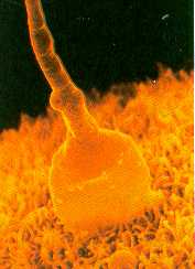
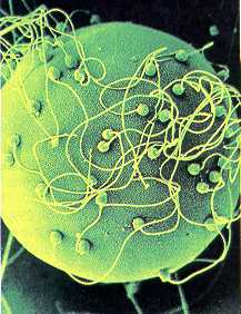
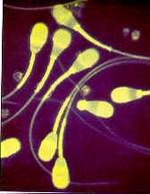
Animals pass on their genetic characteristics to the next
generation via cells called gametes, sperm of the male and eggs or oocytes for the female.
An embryo is formed when an oocyte is fertilized by a sperm (click),
and development of a new animal begins.
About every 20 hours, embryonic cells duplicate their genes and divide,
progressing through 2-, 4-, 8-, and 16-cell stages, etc. The embryo floats freely in the
lumen of the female reproductive tract for the first one to four weeks depending on the
species, and then it attaches to the lining of the uterus, a process called implantation.
The embryo is termed a fetus when recognizable organs form such as the brain and heart.
The sperm, the oocyte before fertilization, and the embryo before
implantation all can be removed from the reproductive tract for various biotechnological
purposes without damaging them. This has led to the development of such techniques as
artificial insemination (A.I.), in vitro (outside the body) fertilization, and
embryo transfer (E.T.), which is the replacement of the embryo into the female
reproductive tract for gestation to term.
Availability of Gametes and Embryos
Nature has gone to great lengths to insure that animals reproduce. For
example, each male of most farm animal species produces trillions of sperm each year, yet
under natural conditions sires only a few dozen offspring per year. Fertilization of each
ovum requires only one sperm. Female farm animals usually produce a few offspring (with
swine a few litters) in their lifetime, yet their ovaries contain hundreds of thousands of
oocytes. The unused oocytes degenerate within the ovaries at a rate of several dozen each
day.
One major principal of biotechnology is to take advantage of the huge
numbers of sperm and oocytes from genetically superior animals that ordinarily would be
wasted by degeneration of oocytes and loss of sperm in urine.
Characteristics of Gametes and Embryos
|
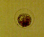
The oocyte is the largest cell in the body, but a microscope
still is required to observe it because it is only 1/200 of an inch in diameter. The sperm
is one of the smallest cells in the body, about 1/6000 of an inch in diameter. By the end
of the first week of development, the embryo grows to more than 100 cells, but it remains
about the same size as the oocyte at fertilization. Thus the embryonic cells get smaller
and smaller during the first cell divisions.
Gametes and embryos are quite resilient to manipulation in vitro provide
that a proper environment is maintained. Sperm are kept in vitro for up to
several days, and we are slowly learning to keep embryos healthy in vitro
throughout the pre-implantation period.
Biotechnology Techniques for Gametes and Embryos
Recovery and Transfer. Sperm are collected with a device called an artificial vagina and/or a rectal electro ejaculator. Depending on the species and other factors, oocytes and embryos are collected and transferred to less valuable females for gestation.
Superovulate the donor cow with various hormones.
Artificial inseminate (A.I.) five days after initiating superovulation.
Non-surgical recovery of embryos(Click) (6 to 8 days after A.I.).
Foley catheter for recovery of embryos.
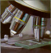
Microscope - Isolation and classification of embryos.
Storage Tank - Storage of embryos indefinitely in liquid nitrogen or one day at room temperature.
Transfer of embryos to recipients surgically or non-surgically.
Pregnancy diagnosis by palpation through the rectal wall 1-3 months after embryo transfer.
Birth (9 months after embryo transfer).
Listed below are the steps taken for cattle:
Cryopreservation
|
|
One of the most useful
biotechnological procedures for both sperm and embryos is cryopreservation. Cooling to the temperature of liquid
nitrogen (-320 degrees F) is done in a medium containing chemicals called cryoprotectants.
Sperm and embryos can be kept in suspended animation at this temperature for decades, and
then thawed, resulting in normal offspring. Storage may be successful at this temperature
for hundreds, if not thousands of years.
Cryopreservation provides great flexibility for various applications.
Semen can be stored, eliminating the need to have males in proximity when semen is used.
Semen and embryos can be sent from country to country inexpensively.
Strains of animals that are no longer of economic importance can be kept
frozen at low cost for a future genetic resource, and animals that have been dead for
years can become genetic parents.
Screening for Genetic Diseases
|
Another option with embryos is to examine several cells for genetic characteristics such as sex and chromosomal abnormalities. This is comparable to amniocentesis which is the surgical insertion of a hollow needle through the abdominal wall and into the uterus of a pregnant female to obtain fluid for the determination of the sex of the fetus or chromosomal abnormality. Knowing the sex of the embryo can be useful for commercial as well as experimental purposes.
Splitting Embryos
|
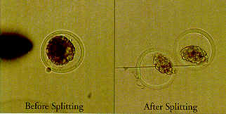
One of the most exciting techniques is the
microsurgical division of embryos into two, three, or four groups of cells. The relatively
simple procedure frequently results in identical twins, and occasionally in identical
triplets or even quadruplets. This should not be to surprising because identical twins occur quite often in nature--and even identical triplets in rare
instances.
Splitting bovine embryos to produce identical twins is fairly simple, and
success rates are excellent. Thousands of calves have been produced from split embryos.
The main reason for using this procedure is a commercial one. Pregnancy rates from unsplit
embryos frequently are about 65 percent; with split embryos they usually are about 50
percent per half; however, since there are two halves, the net result is 100 percent
pregnancy rate on the average, or about one and one-half times as many calves as with
unsplit embryos.
Of course, in many cases only one calf is produced after splitting an
embryo, and in some cases neither half develops. When both halves result in calves, these
identical twins are useful for experimental applications. For example, in certain
nutrition studies about only one-third as many animals are required for valid experiments
if identical twins are used.
Cloning
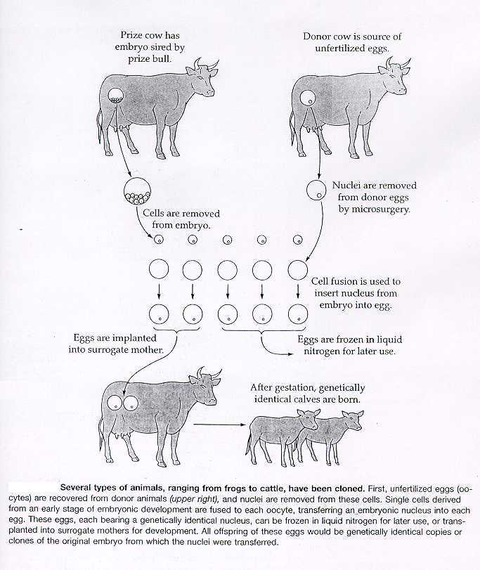 click to enlarge |
In some respects, dividing embryos
to make identical twins, triplets, and so forth is a form of cloning. The resultant
animals are genetically identical and, in fact, might be even more identical than
offspring produced by transplantation of nuclei into oocytes.
Theoretically, more than four identical copies could be produced by
nuclear transplantation, since each cell within the body of an animal has the same genetic
material even though cells of different tissues use different parts of the genetic
information available. While it should be possible to produce large sets of
genetically identical animals eventually, the only practical current method for cloning is
dividing embryos.
In Vitro Fertilization
The Future? It's Here and Now!
Today:
Older Cows: In the past, advanced age caused many cows with genetic merit to be eliminated from the breeding pool. Today these valuable old females may be able to generate a low-risk harvest of immature oocytes, or eggs.
Problem Cows: Females of all breeds and ages may have reproductive difficulties due to environmental causes: ovulatory failure, oviductal transport failure, disease/degeneration of the uterus, and non-responsiveness to stimulatory hormones. Even with these conditions, many cows can be managed to produce ovarian follicles which contain recoverable oocytes.
Healthy Cycling Females: Donor females can be enrolled in an in vitro fertilization program simultaneously with the classical multiple ovulation and embryo transfer. By combining oocyte retrieval and the in vitro fertilization program between rest periods in the superovulatory process, donors reach maximum success.
Terminally Ill Donors: In addition to the use of ultrasound to retrieve oocytes, ovaries from terminal donors can be processed. The ovaries need to be in warm (37 degree C) phosphate buffered saline PBS or saline in a Styrofoam cooler. Use counter-to-counter air service or drive the ovaries to an In vitro fertilization center within 6-8 hours.
The Future:
Young, Prepuberal Heifers: Viable oocytes are produced long before a reproductive cycle can be detected. The net result of this usage is a shorter generation interval ... measured in months instead of years. More offspring are produced from a valuable heifer!
Pregnant Cows and Heifers: During the first trimester (3 months) of pregnancy, many cows continue to produce viable oocytes. The collection process used does not interfere with the developing embryo, so the normal pregnancy proceeds on schedule. Females on this regimen can produce a vastly increased number of offspring in their lifetime.
- Animal Health Livestock and Pets, Yearbook of Agriculture
- New Options in Embryo Transfer, TransOva Genetics, Sioux Center, Iowa 51250
- Supplemental Handout, Carol A. Bluhm, Ferris State University
E-mail for More Information.
This page was last updated April 15th, 1999 © by Carol A Bluhm, Mapleview Farms